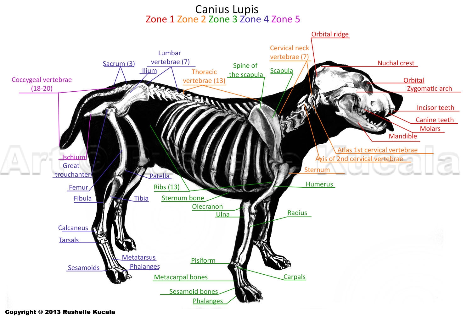
Dog Skeleton Anatomy by TheDragonofDoom on DeviantArt
The Anatomage Dog is the first highly detailed dog anatomy atlas that comprehensively features internal organs, including vascular systems and muscular-skeletal structures. Originating from real dog data, the Anatomage Dog exhibits the highest level of anatomical accuracy. All of its volumetric 3D and individual structures are segmented, users.
Canine Skeleton Poster Clinical Charts and Supplies
An overview of the anatomy of the canine skeleton.Follow on twitter @ https://twitter.com/PerkyVetInstagram: Perkydvm

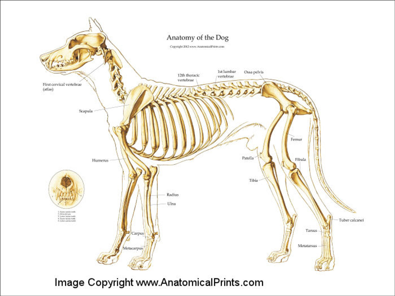
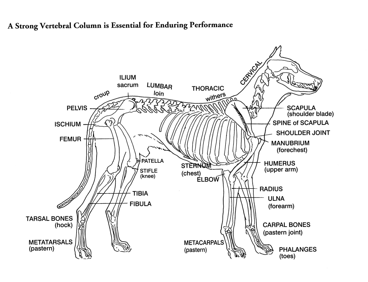
Dog skeleton with major bone elements labeled (Davis, 1987, p. 54
Cross-sectional anatomy - Sagittal: Nasal cavity, Tongue Cranium - 3D - Dog - Anatomy atlas Veterinary anatomy - Dog: Bones - 3D - Dorsal view Rostral view - 3D: Incisive bone, Nasal bone, Maxilla,Frontal bone Labrador - Regions of face: General Anatomy
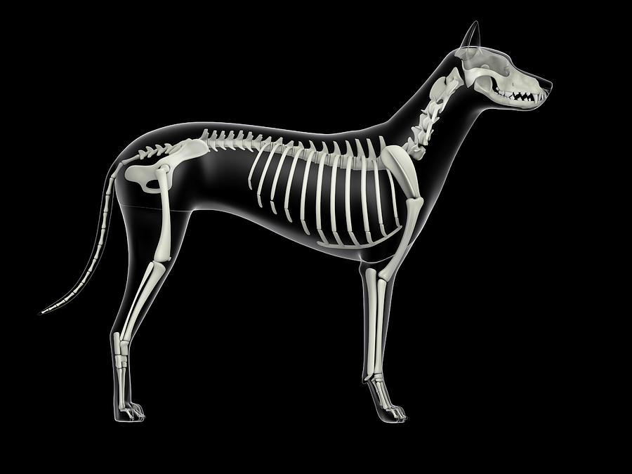
Skeletal System Of A Dog, Xray Side Photograph by Stocktrek Images
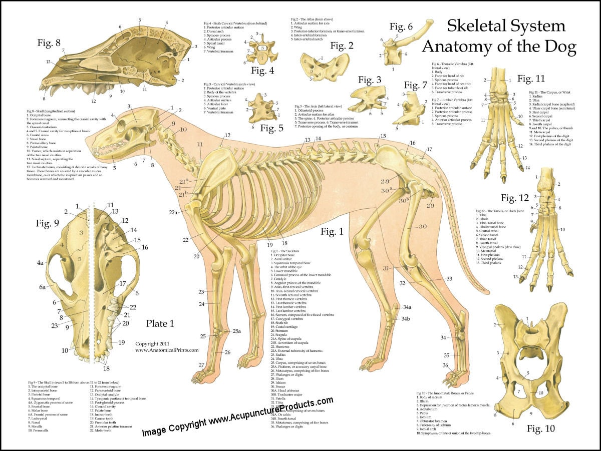
This comprehensive Canine Skeletal Anatomy Poster / Worksheet (Interactive & Printable PDF) is a great revision tool for veterinary students, animal lovers or individuals studying dog anatomy. It is anatomically accurate, vibrant and highly detailed, making it easy to recognise and learn the bones of a canine. It can be used as an offline study.
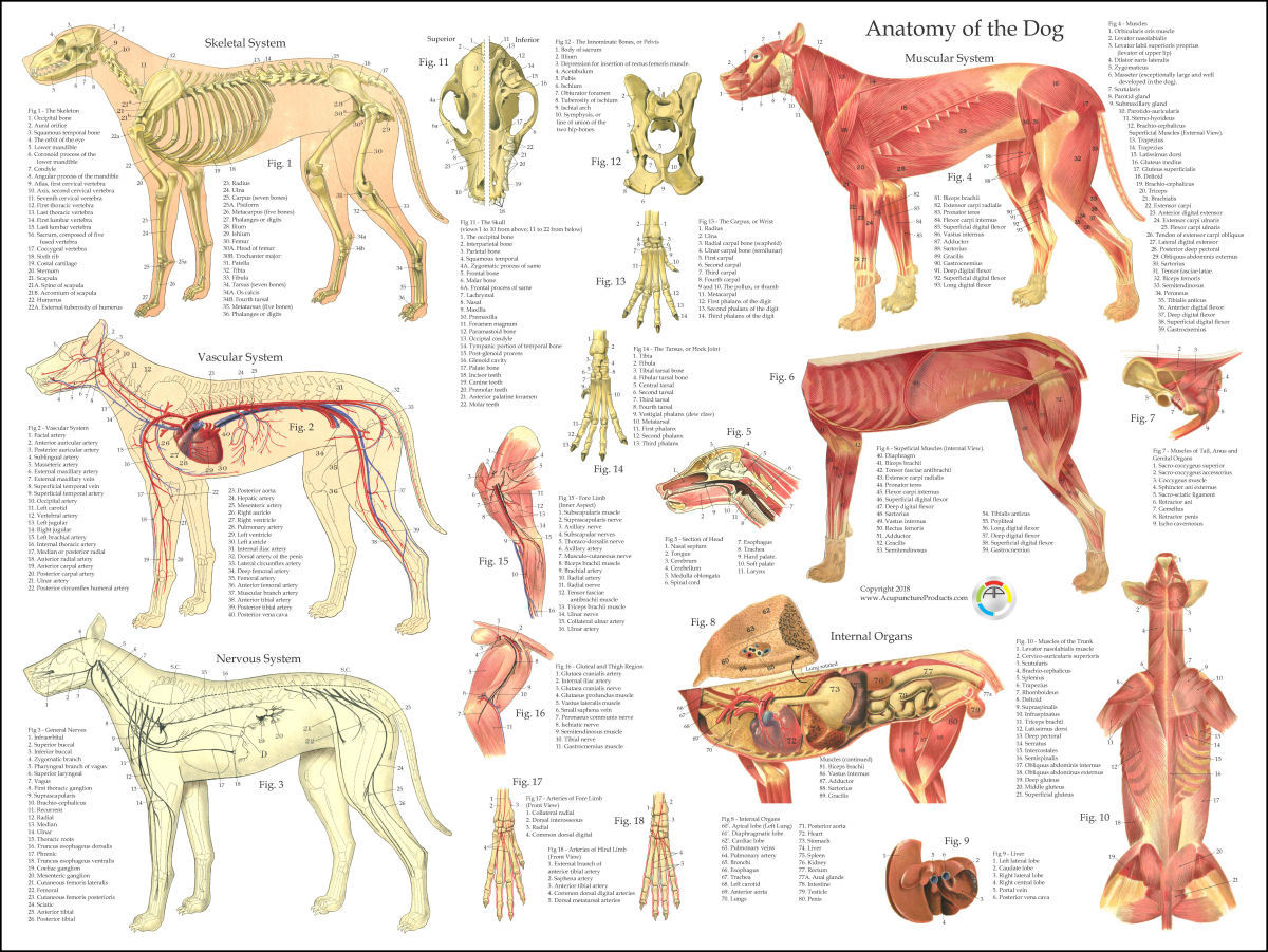
A Visual Guide to Dog Anatomy (Muscle, Organ & Skeletal Drawings) All
It provides information about a dog's skeletal, reproductive, internal, and external anatomy, along with accompanying labeled diagrams. After mating, dogs experience something called a copulatory tie, wherein they remain in the coital position. The male dog dismounts the female at this time. The dogs can remain in this position from a few.
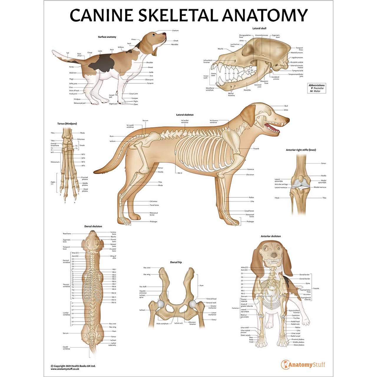
Canine Skeletal Anatomy Laminated Chart Dog Skeleton Poster
The cat has a small coronoid fossa medial to the radial fossa that accommodates the coronoid process of the ulna during elbow joint flexion.; The cat has a supracondylar foramen near the medial condyle allowing the passage of the median nerve and brachial blood vessels.; There is an intermediate tubercle between the greater and lesser tubercles in the horse's intertubercular groove.

Anatomy of dog skeleton with labeled inner bone scheme vector
Come and check all categories at a surprisingly low price, you'd never want to miss it. Awesome prices & high quality here on Temu. New users enjoy free shipping & free return.

Anatomy of a male dog crosssection, showing the skeleton and internal
Components of the Musculoskeletal System in Dogs. Bones provide rigid structure to the body and shield internal organs from damage. They also house bone marrow, where blood cells are formed, and they maintain the body's reservoirs of calcium and phosphorus. Old bone tissue is constantly replaced with new bone tissue in a process called.

FileDog anatomy lateral skeleton view.jpg
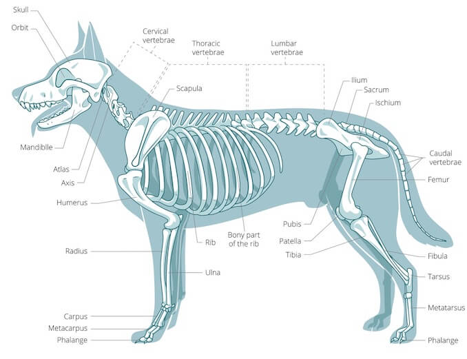
The skeletal system is the 'framework' upon which the body is built - it provides support, protection and enables the animal to move (Fig. 3.1). The joints are considered to be an integral part of the skeleton. The skeletal system is made of the specialised connective tissues, bone and cartilage. Fig. 3.1 The skeleton of the dog showing.
Dog Muscle Skeletal Veterinary Internal Anatomy Poster 18 X 24
Dog anatomy comprises the anatomical studies of the visible parts of the body of a domestic dog.Details of structures vary tremendously from breed to breed, more than in any other animal species, wild or domesticated, as dogs are highly variable in height and weight. The smallest known adult dog was a Yorkshire Terrier that stood only 6.3 cm (2.5 in) at the shoulder, 9.5 cm (3.7 in) in length.

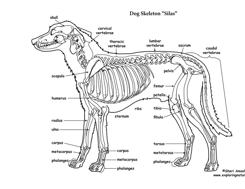
Dog skeleton 101 Dog Anatomy Bones Animal Hackers
In the big picture, the dog skeleton is made of two basic parts: axial and appendicular (limbs). Axial skeleton = head (skull) + the spine (made of vertebrae) + ribs + sternum. Appendicular = forelimb bones + hindlimb bones. The axis of the dog skeleton is composed of the skull, spine (made of vertebrae), ribs and sternum (made of sternebrae).
Helen King on Structure Evaluation Susan Garrett's Dog Training Blog
Skeleton of the dog. Axial Skeleton. = the Head and spinal column. Head - made up of around 50 bones. Skull - occipital crest along top, occiput - rear point of skull zygomatic arch - under and around the base of the eye. Muzzle - nasal crest (largely cartilaginous) top of nose. - maxilla - top jaw. mandible - bottom jaw.

Labeled atlas of anatomy illustrations of the dog Bones Skeletal
Fast and Free Shipping On Many Items You Love On eBay. Looking For Skeleton Bones? We Have Almost Everything On eBay.
Dog Skeletal Anatomy
Dog Skeleton Anatomy. With the large range of breeds and dog sizes, despite their difference in appearance, it might be surprising to hear dog anatomy is generally the same with regards to physical anatomy and characteristics. Dogs have a skeletal system. However, dogs don't have a collar bone, unlike humans; providing a larger.

Dog Anatomy Dog Skelton
Dog skeleton anatomy. The bones of the dog skeleton anatomy serve to support and protect the visceral organs. Again, all the bones of the dog skeleton provide lavers for muscular action. You will find a total of three hundred and twenty-one (321) bones in the dog skeleton. The axial skeleton contains one hundred and thirty-four (134) bones.
Dog Skeletal Skull Anatomy Poster 18 X 24 Etsy
Dog skeleton. As with any vertebrate animal, the skeleton of a dog has the function of supporting the body for movement and protecting its internal organs. We can divide the canine skeleton into three main sections: Axial skeleton: skull, spine, ribs and sternum bones. Appendicular skeleton: bones of the extremities.